About This Project
This project came about when we were looking for co-ops or internships for the summer of 2016. While we were in the midst of applying for traditional co-ops and internships, Nick came up with the idea of continuing and expanding upon a project that we had completed in a class instructed by Josh Shagam. Initially, we applied for a grant to make a start-up company with this idea but were not selected. Unwilling to let this setback keep us down, we turned to our department head, Michael Peres. Initially, he concluded that we should table this project and work on it as a group capstone instead of as a summer co-op, but we weren’t convinced that it should be a capstone. After meeting with him several times, as well as Lisa Vasaturo, we finally got the go-ahead to pursue our co-op over the summer.
The concept for this project started with educational posters being made and printed. The posters included a high quality stitched image of a full specimen. They would also have call outs that had higher magnification images of areas of interest within the specimen, as well as written information about the specimen. These posters were large and somewhat cumbersome, so they were digitized and put on the web. This not only made them less bulky, but also allowed for interactive features to be added. Initially, users could only zoom in and out of the images. As the posters continued to be made for the web, however, more and more features could be added. The information could be placed on a separate layer and areas of the specimen could be highlighted on other layers so that as a user clicked around and scrolled over elements more information was made available. From that, we made a website and started imaging our own samples to populate it. We also created a way to test the color accuracy and image system quality of the microscopes. This allowed us to get better images than ever before with better consistency. Our collection of specimens allows users to view high quality images of specimens and interact with them to learn more about them. Users can access the specimens from mobile devices, as well as laptops.
Over the summer we have worked very hard to bring the Virtual Microscopy Lab to fruition and we hope that it will be a useful tool for learning, as well as interesting and fun to use.
Our Team
We made VML possible!

“Nick is a senior from White Plains, NY, on track to graduate with a degree in Photographic Science in May 2017. As project leader for VML, he supervised the creation of the website, as well as programming the customized scripts used to create the efficient image viewer. In his spare time, Nick enjoys working on his Jeep Wrangler, developing software, taking photos, and conducting experiments.”
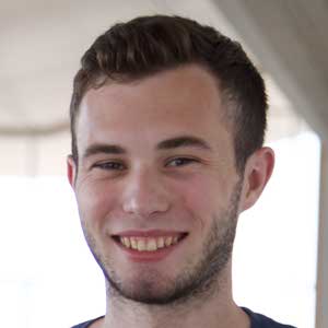
“Justin is a senior in RIT's Photographic Science program, from Boston, Massachusetts. His responsibilities in the project include developing image processing routines and designing calibration methods to ensure color constancy. In his free time, he enjoys cars, film photography, designing computers, and building drones.”
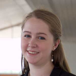
“Kirsten is a fourth year student from south central PA. She plans to graduate with a Bachelor’s degree in Biomedical Photographic Communications with a minor in Japanese from Rochester Institute of Technology. In her free time, Kirsten enjoys cooking for herself as well as friends.”
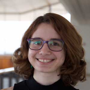
“Teresa Kugler is a third year student from Brooklyn, NY. Her school plan is a double major in Photographic Sciences and Applied Statistics. She is also planning on minoring in music performance. On her free time, Teresa enjoys making art as well as reading in a quiet space.”
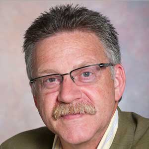
“Michael Peres joined the teaching faculty of the Rochester Institute of Technology School of Photographic Arts & Sciences in 1986. He is a professor of biomedical photography and teaches photomicrography, biomedical photography and other related applications of photography in science. He served as the chair of the biomedical photographic communications department from 1989 until June 2010 when he was appointed as associate administrative chair of the School.”
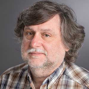
“Steve is Founder and Principal of Acolyte Color Research, a firm specializing in Digital Color Imaging Science. He also served 10 years as a member of the graduate faculty of the School of Printing Management and Sciences at RIT, where he taught courses in image reproduction theory, ink and paper science, research methods, and color measurement. Professor Viggiano was a multiple nominee for RIT's Eisenhart Award for Excellence in Teaching. Before founding Acolyte, Steve had over 14 years' professional experience in Digital Color Imaging Science at RIT Research Corporation.”